
Application of portable ultrasound in emergency treatment
Application of portable ultrasound in emergency treatment
Clinical application of emergency ultrasound
With the continuous development of society, ultrasound examination has become one of the indispensable examination means for medical diagnosis. In emergency treatment, portable ultrasound examination has a wide range, high accuracy, fast inspection speed, non-trauma and no contraindications. Repeated examination can quickly screen patients in any scenario, win precious rescue time for patients with severe fatal trauma, and make up for the shortage of X-rays. Mutual verification with X-ray examination; The biggest advantage is that emergency patients with unstable circulation or who should not be moved can be examined anytime and anywhere, and there is no scene limitation, which is the first examination method for critically ill patients.
1. Application of portable ultrasound in trauma first aid and acute abdomen
Focus Ultrasound Assessment of Trauma (FAST) : Six points (subxiphoid, left epigastric, right epigastric, left renal area, right renal area, pelvic cavity) were selected for rapid identification of fatal trauma.
01 Detection of acute blunt force or acute air injury in the trunk and free fluid in the abdomen: FAST examination is used for preliminary detection of pleural hemorrhage, and to determine the bleeding site and amount (pericardial effusion, pleural effusion, peritoneal effusion, pneumothorax, etc.).
02 Common injuries: liver, spleen, pancreas injury.
03 Common non-traumatic: acute appendicitis, acute cholecystitis, gallstones and so on.
04 Common gynecology: ectopic pregnancy, placenta previa, pregnancy trauma, etc.
05 Pediatric trauma.
06 Unexplained hypotension and so on require FASA tests.
2. The application of portable ultrasound in heart
Echocardiography is the gold standard in the diagnosis of many heart and pericardial diseases.
01 Pericardial effusion: Rapid identification of pericardial effusion, pericardial tamponade, ultrasound-guided pericardial puncture.
02 Massive pulmonary embolism: Echocardiography can help rule out conditions with symptoms similar to pulmonary embolism, such as cardiac tamponade, pneumothorax, and myocardial infarction.
03 Left ventricular function assessment: Left ventricular systolic function was assessed by rapid scan of left major axis, left minor axis, apical four-chamber heart, and left ventricular ejection fraction.
04 Aortic dissection: Echocardiography can detect the location of the dissection, as well as the site of involvement.
05 Myocardial ischemia: Echocardiography can be used to examine the heart for abnormal wall movement.
06 Valvular heart disease: Echocardiography can detect abnormal valve echoes and changes in blood flow spectrum.
3. Application of portable ultrasound in lung and diaphragm
01 Used to assess the severity of early-middle stage pneumonia, small flaps of pulmonary hydrophilia appear in the lungs - Line B sign.
02 Used in the diagnosis of severe pneumonia patients, both lungs diffuse fusion B-line, showing "white lung" sign, severe cases appear lung consolidation.
03 For the diagnosis of pleural effusion, ultrasound guided puncture drainage of pleural effusion.
04 For the diagnosis of pneumothorax: stratospheric sign, lung point and other signs suggest the possible presence of pneumothorax.
05 Guide the setting of ventilator and observe the situation of lung reexpansion.
06 For diaphragmatic ultrasound applications, guided off-line, differentiating central and peripheral respiratory failure.
4. Application of portable ultrasound in muscle tendon
01 Ultrasound can assess whether the tendon is torn and the extent of the tear.
02 For patients with pain and swelling of the hands and feet, ultrasound can quickly and reliably diagnose tenosynovitis, which helps to improve the quality of care and select the appropriate treatment.
03 Assess joint involvement in chronic arthritis.
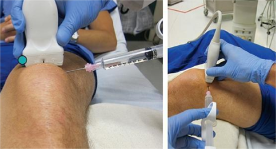
04 Accurately guide tendon and bursae aspiration and soft tissue injection.
5. Application of portable ultrasound in clinical guidance
01 Vascular puncture: visualization of deep vein catheterization, arterial puncture, etc.
02 Guide placement of laryngeal mask.
03 Guided trachea intubation.
04 Joint puncture, nerve block, etc.
05 Guide pericardial cavity, thoracic cavity, abdominal cavity, etc.
06 Cyst, abscess puncture guide, etc.
It can be seen that the application range of portable color Doppler ultrasound diagnostic instrument is extremely wide, and the inspection range is wide, high accuracy, fast inspection, non-trauma, no contraindications, repeated inspection; Portable color Doppler diagnostics have the following important advantages:
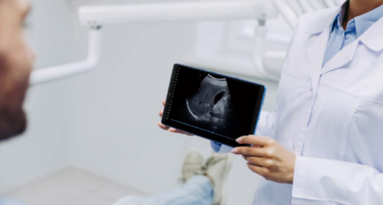
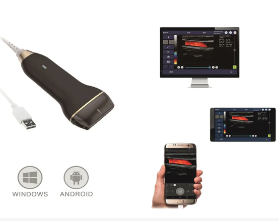
Small and portable, it can be directly carried by hand, which is conducive to medical personnel to quickly carry ultrasound to the medical scene.
The inspection speed is fast, can be repeated, no trauma, no contraindications.
Adapt to a variety of operating environments, including bedside, ICU, emergency, field visits, etc.
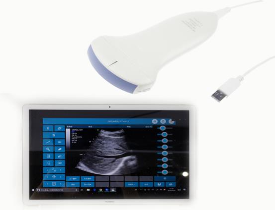
Excellent image quality and support for abdominal, superficial and cardiac probes with a comprehensive range of applications to meet different clinical needs.
Ultrasound interventional therapy, an ultrasound eye carried by the clinician.
The portable color Doppler diagnosis instrument provides a reliable basis for further clinical diagnosis and treatment, and realizes that the critical patients can complete the bedside cardiac ultrasound examination without leaving the ICU, which greatly improves the diagnosis and treatment level of serious patients.