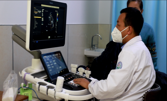
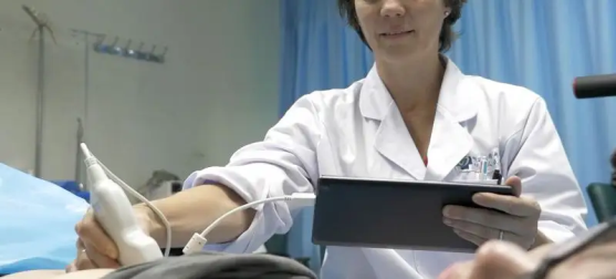
Application range of ultrasound examination
Application range of ultrasound examination
Ultrasound diagnosis of chest and abdomen
1. Diagnosis of thoracic diseases. Diagnosis and differential diagnosis of masses including thymic cysts, thymomas, teratoma and malignant teratoma, lymph node tuberculosis and malignant lymphoma (lymphosarcoma, Hodgkin's disease) in the anterior superior mediastinum; Pulmonary emphysema, atelectasis, pulmonary abscess, and substantial space occupying lesions of the lung (lung cancer); Pleural effusion, empyema, pleural tumor and other lesions.
2. Ultrasonic diagnosis of digestive system organs. There are mainly liver, biliary, biliary system, gastrointestinal, spleen and pancreatic diseases. Such as common diffuse liver disease (hepatitis, cirrhosis, liver fluoriasis, fatty liver, etc.), liver abscess, cyst and hematoma, liver hydatid disease, liver benign and malignant tumors (liver hemangioma, primary liver cancer, metastatic cancer, cholangiocarcinoma); Biliary inflammation, biliary calculus, biliary ascariasis, biliary tumors (gallbladder cancer, extrahepatic cholangiocarcinoma); Acute and chronic pancreatitis, pancreatic cancer, gastrointestinal cancer, intestinal obstruction, intussusception and other diseases.
3. Ultrasonic diagnosis of genitourinary system. These include the kidneys, adrenal glands, bladder, prostate, urethra and scrotum. Renal or ureteral calculi, renal failure, renal atrophy, renal hematoma, cysts, tumors of the kidney and adrenal glands (renal cell carcinoma, nephroblastoma, erollocytoma); Bladder calculi, bladder tumor, prostate hyperplasia, prostate cancer, urethral calculi, urethral stricture; Diseases such as scrotal hematoma, hydrocele, cryptorchidism, testicular tumor and epididymal tuberculosis.
Ultrasound diagnosis of gynecology and obstetrics
1. Diseases of uterus and its accessories (fallopian tubes, ovaries, etc.). Intrauterine device exploration, uterine dysplasia, uterine fibroids, uterine adenomyosis, endometrial hyperplasia, endometrial cancer, follicle development monitoring, endometriosis, teratoma, ovarian serous or mucinous cystadenoma (cancer).
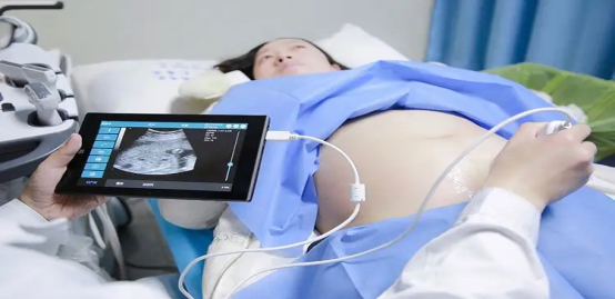
2. Diagnosis of pregnancy uterus. Monitoring of fetal growth, development, amniotic fluid, umbilical cord and placenta in early, middle and late normal pregnancy. Abnormal pregnancies include miscarriage, ectopic pregnancy (ectopic pregnancy), fetal growth and development delay, fetal malformations (anencephaly, hydrocephalus, spina bifida, digestive or urinary system malformations, etc.), placenta previa, placental hemorrhage, amniotic fluid abnormalities, umbilical cord around the neck, and trophotrophic leaf diseases (mole, malignant mole, choriocarcinoma, etc.).
Ultrasonic diagnosis of cardiovascular cavity diseases
Including routine echocardiography, cervical arteriovenous, abdominal arteriovenous, renal artery, limb artery and deep venous system morphological structure, hemodynamic examination. Echocardiography involves placing ultrasound probes in the chest wall and esophagus, conducting numerous section scans of the three-dimensional heart, and comprehensively analyzing the position, morphology, activity and blood flow characteristics of various heart structures, so as to obtain the anatomical, physiological, pathological and hemodynamic diagnostic data of cardiovascular diseases. In recent years, the development of intraesophageal ultrasound, intravascular ultrasound and three-dimensional cardiovascular ultrasound imaging technology has further broadened its application range and greatly improved the diagnostic sensitivity and specificity.
1. Congenital cardiovascular structural abnormalities. Such as atrial deficiency, ventricular deficiency, Fallot III, quadrilateral disease, patent ductus arteriosus, endocardial pad defect, transposition of great arteries, pulmonary venous malformation, congenital valve deformity, etc.
2. Heart valve disease. It can make a definite diagnosis of cardiac valve stenosis, insufficiency, valve calcification, prolapse, perforation, ring calcification, vegetative attachment, and developmental malformation of valve.
3. It is applied to hypertensive heart disease, pulmonary heart disease, hyperthyroid heart disease, cardiomyopathy, aortic dissection aneurysm, aortic sinus tumor and rupture, coronary heart disease, cardiac tumor (myxoma, rhabdomyoma, secondary lung cancer, breast cancer, mediastinal tumor) and intracardiac thrombosis.
4. Endometrial lesions, plaque formation or stenosis of carotid artery, abdominal aorta, renal artery, and great artery of limbs; Thrombosis, dilatation and malformation of veins in the head and neck, abdominal cavity and limbs.
Ultrasound diagnosis of superficial organs
Mainly include thyroid and parathyroid gland, breast, eye, testis, scrotum, maxillofacial diseases, as well as some bone, limb muscle joints, subcutaneous tissue fascia lesions, such as hematoma, abscess and tumor. The examination of these organs requires the use of high frequency probes (above 7.5MHz, mostly 10-15MHz), which have good fine structure resolution.
1. Thyroid ultrasound diagnosis; Simple, nodular, and diffuse goiter (hyperthyroidism); Thyroiditis, thyroid tumors (adenomas, cysts, thyroid cancer); Ultrasound diagnosis of parathyroid hyperplasia, cysts, adenomas and parathyroid carcinoma.
2. Ultrasound diagnosis of breast diseases: mastitis, lobular cystic hyperplasia, breast cysts, breast fibroadenoma and breast cancer. Ultrasound is the first choice for the diagnosis of breast cancer because of its noninvasive, simple and easy operation.
3. Ultrasonic diagnosis of eye diseases: the eye and orbit are located on the surface of the human body, the anatomy is relatively simple, the interface is clear, and the sound attenuation is less, which is one of the most suitable parts for ultrasonic detection. It is mainly used for intraocular tumor, cataract, detachment of retina and choroid, intraocular hemorrhage, ocular foreign body or ocular trauma, and intraocular lens implantation before and after monitoring.
4. There are various injuries, inflammation and tumors of muscles, tendons, ligaments, joints and bones.
Interventional ultrasound diagnosis and treatment
As a branch of modern ultrasound medicine, interventional ultrasound medicine is characterized by the real-time ultrasonic monitoring and guidance, complete a variety of puncture, extraction, biopsy, injection treatment and other operations, can avoid some surgical operations, so as to achieve a comparable effect with surgery. Especially in recent years, the ultrasonic guided automatic biopsy technology with automatic biopsy device has improved the puncture efficiency and the quality and quantity of biopsy specimens, and reduced the damage and complications that may be caused by manual operation, with high accuracy and safety.